Getting My Babyecho To Work
:max_bytes(150000):strip_icc()/JoseLuisPelaezInc-17f79a53211940c2bc62cf23bc4185d4.jpg)
For a lot of ladies, ultrasound shows that the infant is growing normally. In some cases, ultrasound might reveal that you and your infant need special care.
A c-section is surgical procedure in which your infant is birthed with a cut that your physician makes in your stomach and uterus. Regardless of what an ultrasound shows, talk to your provider concerning the very best care for you and your baby - heart doppler. Last assessed: October, 2019
Throughout this scan, they will inspect the child is growing in the right area, whether there is even more than one baby and they will certainly also examine your child's advancement until now. This testing is readily available in between 10 14 weeks of maternity and is utilized to examine the possibilities of your child being born with several of these conditions.
Babyecho Fundamentals Explained
It includes a mixed test of an ultrasound check and a blood test. During the scan, the sonographer will certainly determine the liquid at the rear of the baby's neck to identify 'nuchal translucency' - https://www.bitchute.com/channel/b9AwfZqOVru6/. They will certainly after that determine the chance of your child having Down's, Edwards' or Patau's syndrome using your age, the blood examination and check outcomes
Throughout this scan, the sonographer look for architectural and developing abnormalities in the child. During this scan consultation, you might be used screenings for HIV, syphilis and liver disease B by a professional midwife. In some situations, a third-trimester check is recommended by your midwife complying with the results of previous tests, previous issues or existing clinical conditions.
The traditional 2D ultrasound produces flat and described pictures which can be utilized to see your child's interior body organs and assist identify any type of internal concerns. These black and white photos help the sonographer determine the child's gestation, development, heartbeat, growth and size. Some pregnant moms choose to have a 3D ultrasound check due to the fact that they show even more of a real-life photo of the baby.
Rumored Buzz on Babyecho
3D ultrasound scans reveal still photos of your infant's exterior body as opposed to their withins, so you can see the shape of the baby's facial features. 4D ultrasound scans resemble 3D scans however they reveal a relocating video clip as opposed to still photos. This records highlights and shadows much better, therefore producing a clearer photo of the baby's face and motions.
:max_bytes(150000):strip_icc()/191127-ultrasound-trimester-pink-2000-fd089add04f8444e9d7a403933d1994f.jpg)
or (8-11 weeks) (11-14 weeks) (14-18 weeks) (19-23 weeks) or (24-42 weeks) Advised at Optional -, more regularly in some problems This check is done to and to establish an (EDD). A is discovered throughout this scan. A lot of parents choose this check for. Is crucial prior to the blood examination called as (NIPT) to determine the.
A Biased View of Babyecho
Sometimes a may be called for to get and a clearer image. This is usually carried out and occasionally a might be required (baby doppler). Confirm that the infant's heart is existing; To more accurately.
Please see below. It coincides as 19-22 weeks, yet some might be or in the and it might to. Typically this is provided if there are such as spina bifida or if parents are keen to recognize the earlier. These scans may be done, nevertheless a few of the and therefore, a is needed to This check is done typically at.
Unknown Facts About Babyecho

Furthermore, the can be by by an. () The way nearer the is beneficial to. Periodically, an which was previously might be.
Not known Factual Statements About Babyecho
If, these scans might be to. on the searchings for, a might be used. During all the, a 3D scan (of the baby) can likewise be performed. The hinges on the placement of the,,, quantity of and. This consists of, in addition to; This consists of, along with (14-20 weeks).
A scan is essential prior to this test is done.
Babyecho Things To Know Before You Get This
The test can provide beneficial information, helping females and their health-care service providers handle and care for the maternity and the fetus.
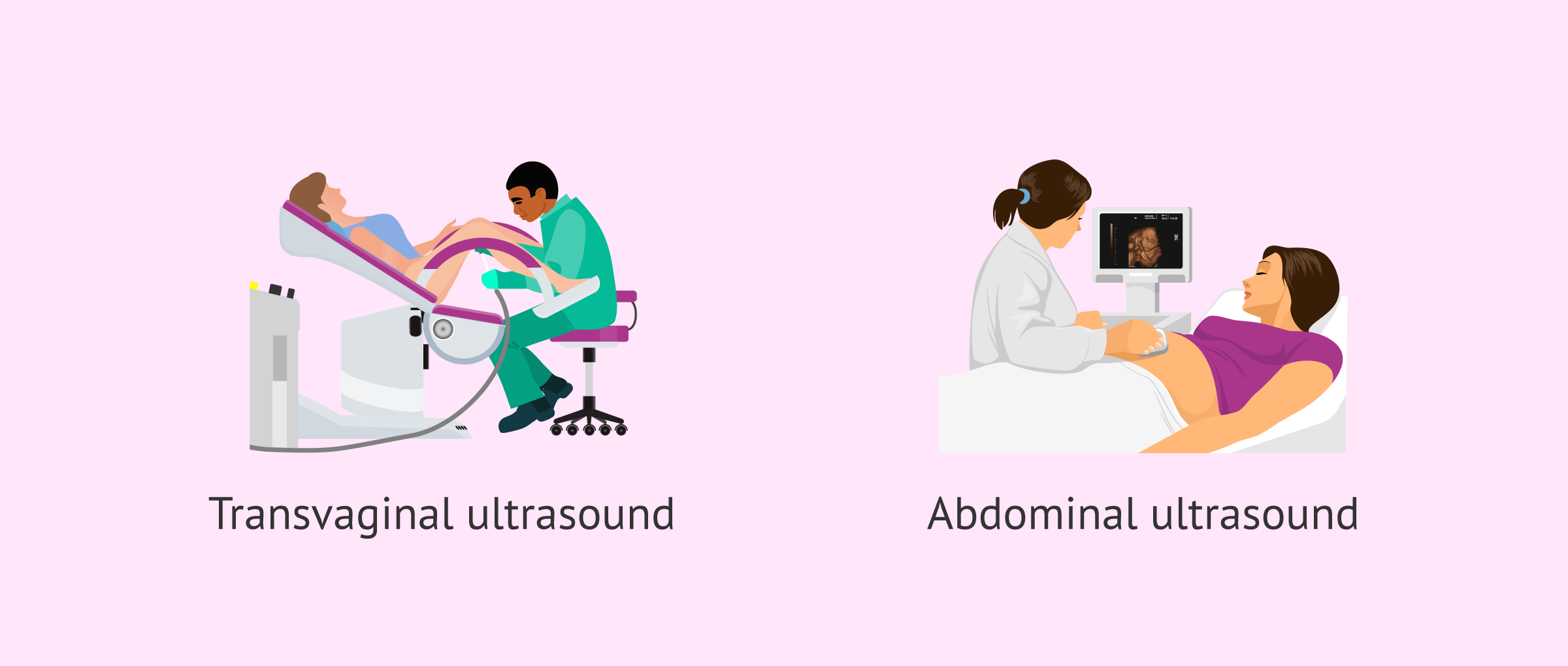
A transducer is put right into the vaginal area and relaxes against the rear of the vagina to develop a photo. A transvaginal ultrasound produces a sharper image and is frequently utilized in very early maternity. Ultrasound equipments are regarding the dimension of a grocery store cart. A television display for seeing the photos is affixed to the equipment (https://urlscan.io/result/2eca35a6-7c7f-485b-bdc6-aaf453125190/).
Comments on “Indicators on Babyecho You Should Know”